

Dr. Abdullah Mahmud
GENERAL DENTIST
University of Detroit Mercy, Class of 2018
Qualities that describe me:
* Always striving to improve
* Motivated to learn the best techniques
* Empathy towards my patients
Accomplished individual clinically and didactically.
Constantly searching for the latest CE, dedicated to patient care, and always striving to provide the highest quality of service.
WORK EXPERIENCE
Friendly Dental Durham, NC: August 2023 - present - Full scope of general dentistry especially molar endodontics, full bony third molars, and crown and bridge. Some implant dentistry (placement and restorative)
Comprehensive Dental Care Rocky Mount, NC January 2024 - January, 2025 - general dental services, primarily focused on a Medicaid patient base. Mostly extractions, restorative restorations, and removable prosthodontic cases (dentures and partials).
Friendly Dental Charlotte, NC: November 2019 to November, 2023 Oral surgery, restorative, and crown & bridge. Extensive experience in removal prosthodontics, oral surgery including impacted third molar teeth and endodontics.
Dr Taylor Family Dental Center Waterford Twp, MI: May 2019 to August 2019: Part-time position focusing heavily on restorative, endodontics, and extractions. Extensive experience working in pediatrics.
Dental Care of the Future Fraser, MI: May 2018 to August 2019: Full-time position where I was involved in the full scope of general dentistry procedures including molar endodontics, extractions, and socket grafts. This office also had a unique method of diagnosing periodontal disease based on microscopic findings.
Drakeshire Dental Care Farmington, MI: June 2018 to April 2019: Restorative & Crown/Bridge focused
EDUCATION
2014-2018
Doctorate of Dental Surgery
University of Detroit Michigan
Detroit, MI
2004-2008
Bachelor of Science in biology
Bachelor of Arts in German linguistics and Translation
University of North Carolina at Charlotte
Charlotte, NC
Portfolio of case completions available upon request in the areas of prosthodontics and endodontics. Skilled in fixed and removable prosthodontics, composite restorations, molar endodontics, simple and surgical extractions, impacted third molar extractions, suturing, and emergency dental services. Socket preservation bone grafting. CEREC experience in fabrication of crown & bridge. Implant restorative experience and some exposure to implant placement. Extensive experience with all patient populations including a heavy exposure to pediatrics and nitrous oxide sedation.
Fluent in German and Arabic.
Minor fluency in Spanish.
North Carolina DDS - License Number 11523
Michigan DDS - License Number 2901022621
ACTIVE LICENSURE:
SKILLS
CLINICAL PORTFOLIO
Four Canal Maxillary First Molar


Four Canal Lower Second Molar


5 canal lower first molar
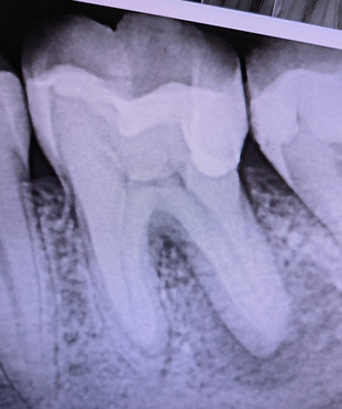


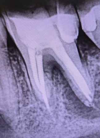
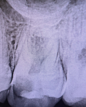
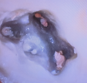
Five Canal Maxillary First Molar
MB, MB2, DB, DB2, Palatal

Five Canal #14 same day root canal therapy finding all five canals (MB, MB2, DB, DB2, P) and core, and crown. Temporary crown shown in this PA radiograph. Wave One Dentsply System.
FIVE CANAL LOWER FIRST MOLAR
Three Mesial Canals
Two Distal Canals


Middle aged female with generalized attrition and habitual edge to edge habit that received a full mouth reconstruction with 28 zirconia crowns. Patient had minimal VDO loss and had a Class I bite with a consistent slide into edge to edge and Class III bite scheme.
Patient had generalized wear on the maxillary and mandibular incisors. You can appreciate the heavy level of wear and grinding that took place with this patient. She was grinding through a bite guard every few years.


After keeping the patient in temporary crowns (Fixed Orthotic temp) for several months, making adjustments, and getting the patient into a habitual Class I bite, we proceeded to eventually get the patient into a perfect Class I occlusion with 28 monolithic zirconia crowns.
Great Class I occlusion that you can appreciate from this video.


Esthetic zirconia in this six unit bridge case in which Teeth #6,7 and 11 served as abutments. Patient was elderly with limited financial means and a complex medical history. Implants were not an option for her.


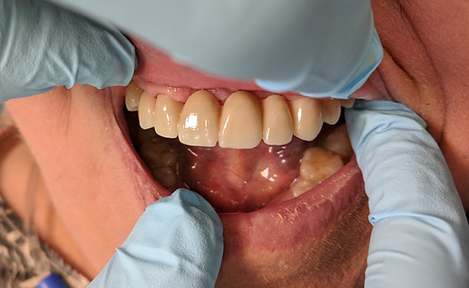
6 unit bridge
#22 - #27


Crowns #7-#10 Good radiographic adaptation. Used Porcelain fused to zirconia material.


Tooth #12
* Root Canal Therapy
* Post & Core
* Crown Lengthening
* Crown
Functional Crown Lengthening Case to expose more tooth circumferentially
Impression taken same day as crown lengthening using Kettenbach Impression Material


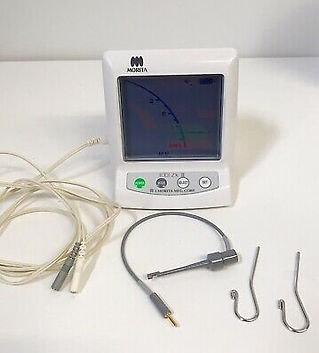
Two cases, both on Tooth #19. These separate cases show that the anatomical apex does not always correspond to the radiographic apex. In both cases, the anatomical apex made a 90 degree turn laterally towards the distal. This goes to show that cases in which gutta percha appears to be short of the radiographic apex may indeed have been filled completely due to the curves that teeth take. Having an apex locator is essential. I prefer the classical Morita apex locator for all my cases. It’s simple, effective, and gets the job done very predictably.


Close up view of the distal root "puff" of sealer showing complete cleaning and shaping of the distal root system

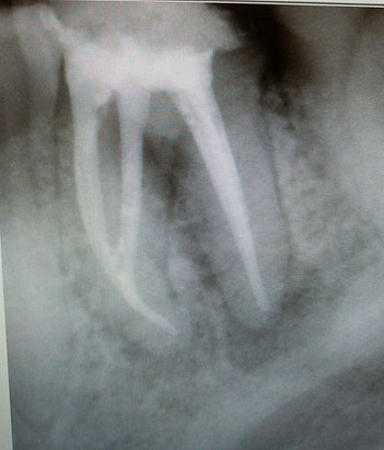
Tooth #19 diagnosis:
Pulpal: pulpal necrosis
Periapical: Acute abscess w/ apical periodontitis
Had to do incision and drainage due to large buccal abscess and large distal periapical infection. Did complete cleaning and shaping, placed calcium hydroxide with cotton pellet and IRM, and put the patient on antibiotics.
Recalled the patient two weeks later and completed the root canal. Four canals with ML joining the MB in the apical third. Patient had complete resolution of symptoms when he presented for delivery of the crown and the tooth went from Class 2 mobility to no mobility after treatment.
Various endodontic cases completed between June 2018 and August 2020.
All cases completed using Vortex Blue rotary and Wave One Gold reciprocating files.


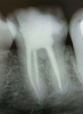


Above Case: Tooth #3 that was extremely calcified and it was difficult to find the canals let alone negotiate them to the apex. This case took 1.5 hours from start to finish and many #8 and #10 hand files, both K files and C files, were used to establish patency. MB, DB, and Palatal canals were cleaned and shaped to full working length. Patient subsequently had a crown fabricated and was completely asymptomatic.


Tooth #31 that required root canal therapy. The patient was a young Asian lady. The tooth actually had THREE MESIAL canals and ONE DISTAL canal. MB, MM, and ML, and Distal. The MM (Middle Mesial) had its own apex along with the other canals.
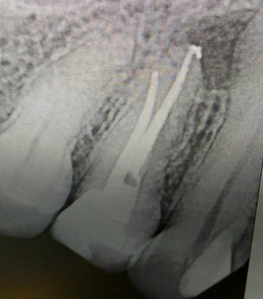
Patient presented with percussion symptoms and no sensitivity to cold.
Diagnosis:
* Pulpal Necrosis
* Symptomatic Apical Periodontitis
Treatment Options:
* Extraction
* RCT/Core/Crown

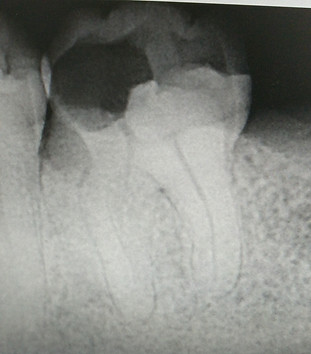
Tooth #19 obturated same day. Tooth had a necrotic pulp and was thoroughly cleaned out with copious sodium hypochlorite. I use the Dentsply EndoActivator to ensure proper introduction of hypochlorite to the entire root canal system.
All four canals instrumented and obturated using the WaveOne Gold primary reciprocating files and obturated using Wave One Gold primary cones with Dentsply Ribbon Sealer. Good prognosis expected.


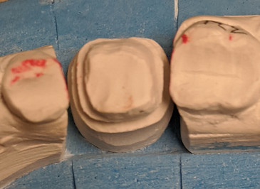

Tooth #30 crown delivery case due to previous MODL restoration fracture

Tooth #2 root canal therapy completed with four canals identified (MB1, MB2, DB, Palatal) . Tooth #1 was extracted at a later date. Tooth #2 was then restored with a full coverage zirconia crown. Distal crown lengthening was needed to allow proper margin placement which was done at the time of the crown preparation. Final crown fit extremely well and patient is happy to keep this tooth in function.

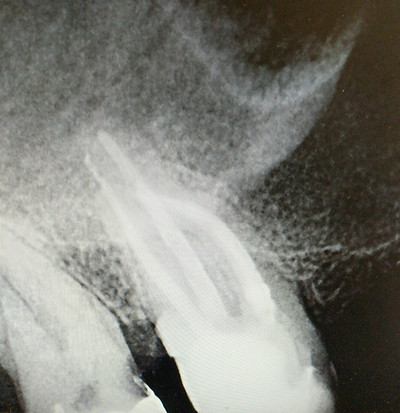
Extractions of multiple teeth as shown in the pre-operative and post-operative Panoramic radiographs. I have done many such cases including full mouth extractions usually in the same visit unless the patient has a medical limit for local anesthetic administration or other factor.


Patient with rampant interproximal decay. The following restorations were placed:
#12: DO
#13: MOD
#14: MODL
#15: MOL
I use a combination of Tofflemire and Garrison or Palodent bands. I use the tofflemire for #12 and #14. Then after contouring the interproximal areas, I use the sectional bands on #13 and #15 being restored simultaneously with three rings on. Excellent contacts areas and embrasure development is possible with this technique.


Tooth #30
Patient sensitive to hot and cold. No percussion symptoms. Previous amalgam with recurrent decay.
Patient presented for crown preparation visit and told pre-operatively he may need RCT. Caries was excavated and mesial pulp horns exposed. Same day RCT, Core, and Crown. Wave One Gold Primary for all four canals (MB, ML, DB, DL) Final crown is a full strength monolithic zirconia crown.
Interesting side point: patient presented two weeks after crown cementation with gingival irritation around the crown only on the buccal. Good contacts and embrasure form. Turns out the patient had very minimal keratinized tissue and the crown was slightly over-contoured facially causing irritation. I used a diamond flame smooth bur to contour the crown and minimize tissue contact. His symptoms disappeared after the facial contouring.


Various Crown Deliveries
Monolithic full strength zirconia crowns

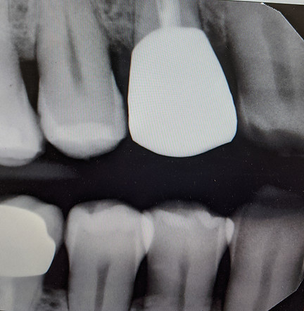
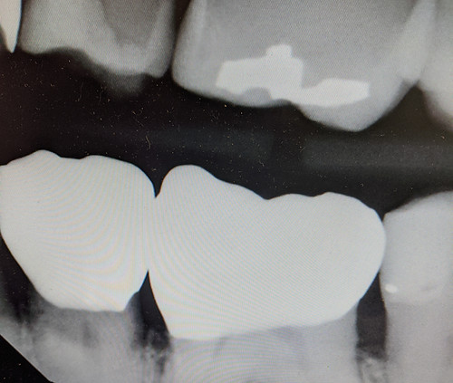


Tooth #6-#11 Anterior Zirconia crowns
Treatment was performed after bringing the patient to an ideal periodontal state. Patient will plan on having a crown placed on Tooth #4 at a future visit and maxillary and mandibular metal framework partial dentures for posterior occlusion. Patient was very satisfied with the appearance and function of her new crowns. Complete black triangle closure was not possible but due to the low smile line it was not something of concern to the patient.


This is the final picture taken 12 weeks after cementation. Patient is extremely happy with the final result in terms of esthetics and function.
As discussed previously, the papilla areas are blunted but that was a pre-operative finding. Patient had a low smile line and was NOT interested in soft tissue grafting.
Below are the radiographs showing good adaptation of these PFZ (Porcelain Fused to Zirconia) crowns on Teeth #6-#11.


Tooth #30 & #31: #31 RCT,Core,Crown & #30 Core,Crown


Esthetic Case Tooth #6- #11 Wax Up used to simulate the finals for the patient for 2 weeks.
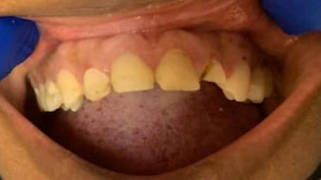


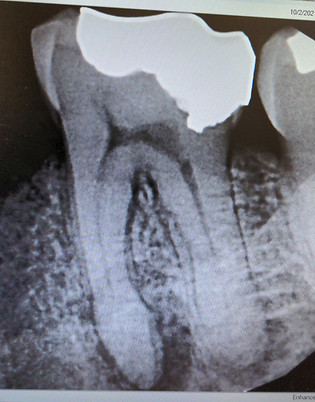

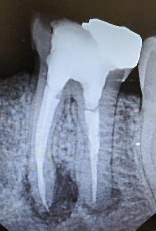
FIGURE A
FIGURE B
FIGURE A depicts #19 that is grossly necrotic. Identified all three canals and placed calcium hydroxide. Figure B is the cone check radiograph with an overextentsion of the distal gutta percha that was trimmed back. Figure C depicts the final radiograph of all three canals.
FIGURE C
Tooth #5: Same Day Root Canal, Core, Crown.
Tooth #5 root canal therapy performed due to decay that reached the pulp. Tooth was root canal treated, then prepared for a full coverage zirconia crown. Temporary crown placed. Final crown cemented two weeks later. Patient very satisfied with functions and esthetics of the tooth.
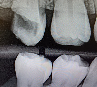
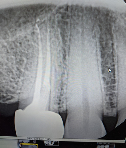

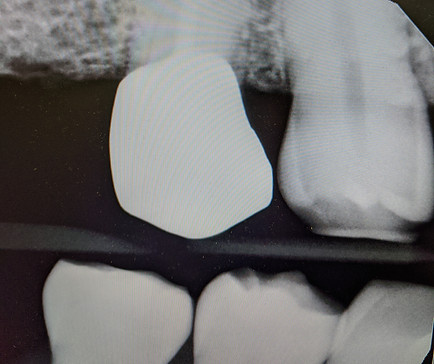
Same day extraction of all remaining maxillary and mandibular teeth. Maxillary third molars were kept in place due to full bony impaction. Remainder of her mouth was non-restorable and extracted.


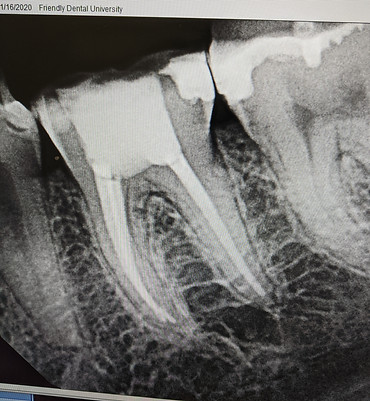
Case to the left:
Tooth #19
Diagnosis: Symptomatic Irreversible Pulpitis
Prognosis: Excellent
3 of canals: Three (MB, ML, Distal)
System: Wave One Gold
Obturation: Corresponding Wave One gold GP points
Sealer: Septodent BioRoot Biocermaic sealer
Obturation Technique: Hydraulic Condensation
Below Case:
Tooth #2
Diagnosis: Pulpal Necrosis
Prognosis: Questionable due to large carious lesion and considerable removal of tooth structure
4 canals identified (MB1,MB2, DB, Palatal)
System: Wave One Gold files
Obturation: Corresponding Wave One gold GP points
Sealer: Septodent BioRoot Bioceramic sealer
Obturation Technique: Hydraulic Condensation
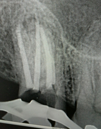
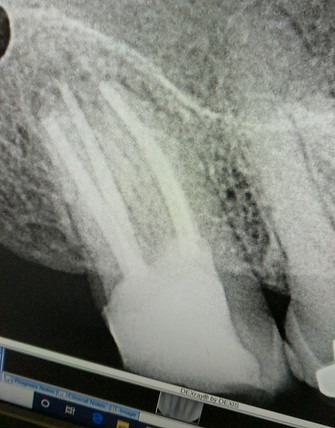

Tooth #30
Diagnosis: Pulpal necrosis
Four canals identified with a torturous and windy pathway to the apex. WaveOne Gold primary files used to clean and shape the two mesial and two distal canals with copious sodium hypochlorite. Patient completely relieved of pain symptoms. Crown preparation will be completed within a few weeks of treatment.
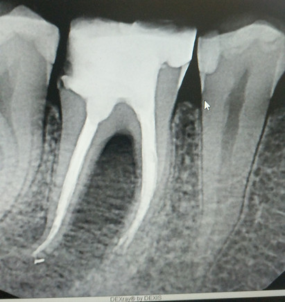

Tooth #18
Diagnosis: Symptomatic Irreversible Pulpitis
Treatment: Root Canal, Core, Crown #18
Patient presented for crown preparation on #18. When patient presented, there were no pulpal or periapical symptoms present. Upon removal of decay, the excavation got close to the pulp. Patient was told there could be a need for root canal therapy after treatment. Upon presenting for crown delivery visit, it was determined that the tooth was undergoing potential signs of irreversible pulpitis. Crown was delivered with temporary cement. Patient presented three weeks later still symptomatic. Root Canal was initiated and completed same day (Three canals that were very vital ---> MB, ML, Distal) Crown delivered with permanent cement and patient was instantly relieved of her symptoms.
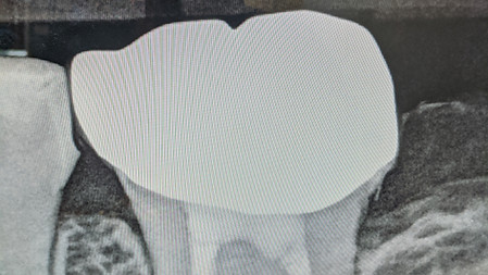

Gingivectomy Procedure
Pre-op, Immediate Post-op, 1 week post-op
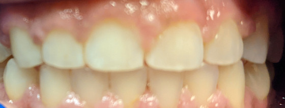


#8,#9 Custom Shade
Base Shade 1M1
Beautiful result with custom shading #8,9 to the incisal edges of #7 and #10.

Tooth #19
Large carious lesion on the buccal. Was able to remove carious lesion completely, rebuild the wall, and do the root canal. Patient came 1 week later and had the crown preparation completed.
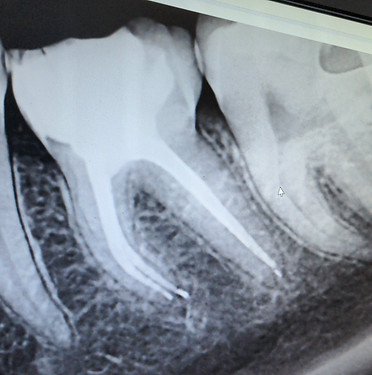

Tooth #3 with four canals. MB2 joined the MB1 in the middle third. Excellent cleaning and shaping using WaveOne Gold rotary system.
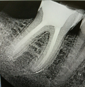
Tooth #30 four canals identified and cleaned and shaped to perfection as can be demonstrated by the slight sealer puffs at the apices of both roots.

Third Molar Extraction Cases
Case #1: #32 mesio-angular impaction. Acute pain resulting in pressure sensation on #31 and extreme pain upon palpation of the gingival tissue above #32. Successfully removed tooth in 15 minutes.

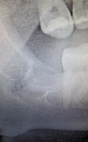
Case #2: Tooth #17 full bony impaction. Successfully removed with buccal, mesial, and distal bone relief.
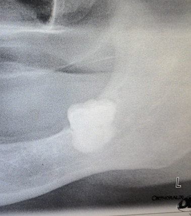

Case #3: Extraction of #1,16,17, and 32. #17 required a good flap to be raised, buccal bone gutter trough, and adequate luxation to allow complete removal of the tooth.
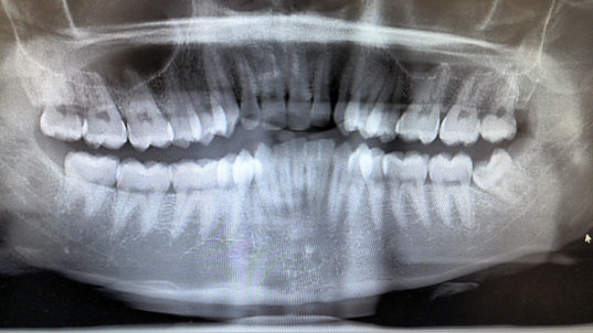

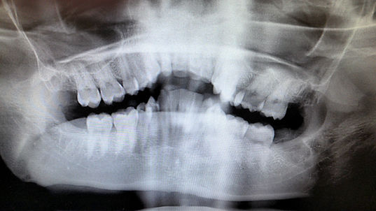
Horizontal Third Molar #32 Extraction and #1 extraction


Apicoectomy
#23 with a lesion that did not heal and patient had symptoms a year later.
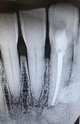
Apico surgery done successfully with complete resolution of symptoms.


